Would you like to begin by introducing yourself?
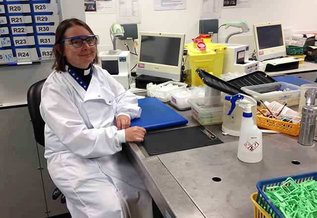
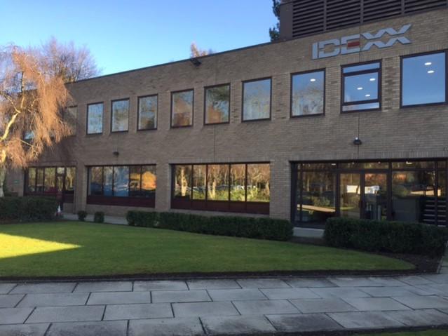
I‘m the Reverend Dr Jenny McKay, a veterinarian by training and with a PhD in Veterinary Neuropathology, and my European and Royal College qualifications in Veterinary Pathology. I am currently Head of European Anatomic Pathology at IDEXX Laboratories Ltd. This is an international company which supports veterinary healthcare globally. Our main UK lab is based in Wetherby, North Yorkshire and I am usually based here 2-3 days a week, although since the COVID pandemic, I have been working virtually from my home in Cheshire.
Could you tell us what sparked your interest in digital pathology in the first place?
I worked for many years in the pharmaceutical industry where I had seen the benefits of digital pathology in terms of quantitative image analysis for animal models of disease.
When I moved back to veterinary diagnostic pathology in 2013, it was obvious that digital pathology could help us in that field too. It could enable us to work with pathologists from all over the globe and produce high quality images without the paperwork and posting back and forwards between individuals for second opinions.
What’s the setup for the IDEXX team – how many people are in the team and what are their job roles?
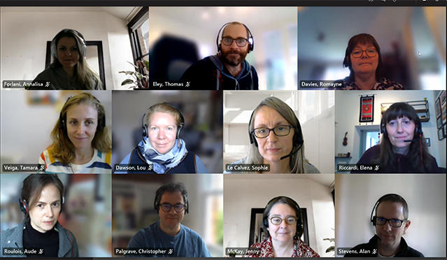
In the UK Team, I work closely with my Deputy Head Dr Sophie Le Calvez, and two Team Leaders, Drs Lou Dawson and Tamara Veiga Parga. Each of them has a team of 6-7 veterinary pathologists. Everyone is reading predominantly small animal biopsy cases which come in from veterinary practices and hospitals across the UK and beyond.
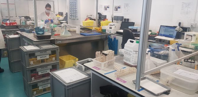
We process upwards of 7500 histopathology samples for pets each month which is a huge volume of cases. There is nowhere else where you could see such a range of cases in canine and feline medicine.
We always have a pathologist on duty each day (called the POD) who deals with pathology phonecall queries from clients and the highly urgent cases. Prior to COVID, we always had a trim pathologist who would deal with specific difficult cases in the histology lab, but we now do a lot of these consults via webcam. Some of the team have special interests such as undergraduate or postgraduate webinar training sessions, and I always encourage team members to publish in high impact scientific journals since we have such a huge bank of animal samples at IDEXX.
Is there anything that you think is particularly unique to your Lab?
We process upwards of 7500 histopathology samples for pets each month which is a huge volume of cases. There is nowhere else where you could see such a range of cases in canine and feline medicine.
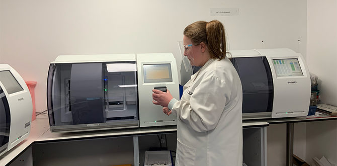
We have a fully automated system where all the samples are barcoded on arrival and we also have some members of the team working from New Zealand enabling 24 hour reporting of cases.
How do you encourage effective collaboration amongst your team and the technical staff?
It is easy to share cases digitally and discuss cases in real time. We also run regular webinar sessions with the technical staff where we present, for example, interesting cases, why we need to use special stains and immunohistochemistry for diagnostics, and the importance of providing good trim notes as the technical staff are really “our eyes” in this respect.
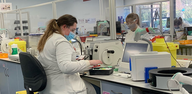
We like to highlight specific animal cases as it reminds everyone that we are working with real animal tissues, not just impersonal bits of tissue. Over the last couple of years we have also introduced “shadowing sessions” where 2-3 technical staff at a time sit with a pathologist for half an hour to observe their work. It really helps build relationships between the pathologists and the technical staff and we could highlight points like how if a section is folded it makes all the difference to interpretation, and this is why we ask for re-cuts.
Could you describe an average week for you and your team?
The main thrust of the team’s work is reporting on canine and feline biopsy cases. However, in order to keep us up-to-date we also have 1-2 training sessions each week where specific types of cases are discussed and reviewed.
Every Friday we also have a Journal Club where we take it in turns to discuss a recent publication.
For example, Dr Le Calvez has a specific interest in lymphomas and Dr Chris Palgrave also presents regularly on skin and mammary cases. Dr Elena Riccardi is an expert in gingival pathology so we are always keeping abreast of new developments and nomenclature. Every Friday we also have a Journal Club where we take it in turns to discuss a recent publication. Second opinions are triaged to those senior pathologists with a special organ interest and if there is still a differing opinion then I will be pulled in! Those team members involved in management, like myself, obviously spend much less time reading cases and we are involved in inter-departmental projects and steering groups.
In normal times we would travel and meet colleagues across the globe, but that has changed recently, and probably will change forever. We generally work between 9.00am and 6.00pm and one or two pathologists will also cover Saturdays.
What are the main advantages of working digitally?
The main advantages are in being able to access cases easily, share cases in real time with colleagues all over the globe, and of course it aids in recruitment. Veterinary pathologists are quite rare and may not want to move their home location, so being able to offer people virtual reading from home is a huge advantage. During COVID, we have appreciated the technology so much, and our service to clients continued in an uninterrupted manner.
How has COVID-19 affected your work? What steps have you taken to overcome the challenges?
Because we already were fully digital and working totally or almost completely from home in any case, the COVID impact has been small.
The main aspect missing though is the physical face-to- face contact with our histology and pathology operations teams and each other. We have maintained our training webinars for technical staff and have had a small number of “coffee” meet ups virtually to keep up team morale.
I always check in with each member of staff on a 1:1 basis every 6 weeks as I feel it’s really important for the pathologists to know that they are not forgotten and it helps me understand the issues some are experiencing with childcare management and isolation.
Could you tell us about an interesting case, when working digitally has made the difference to practicing veterinarians?
With having digital pathology we include images in our reports. This is very helpful when perhaps further surgery has to be performed if margins are too close initially. It really helps the vets explain to clients why re-surgery has to take place.
It is so easy to place images in reports now that we’re not reliant on the slow process of taking photos with a microscope and uploading them. I know that these images really help vet-pet owner discussions on further treatments.
What do you see as the barriers to the further integration of digital techniques into pathology and what is preventing more pathologists from using digital pathology in their work?
At IDEXX, we are fortunate to provide diagnostic services to veterinary hospitals and clinicians. IDEXX invests in the latest technology that will be of benefit to providing the best and most reliable results for our clients. I know in other businesses that implementing digital pathology would be a difficult financial investment.
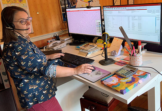
Our digital system has been thoroughly validated and I think more pathologists should take the plunge and realise that the resolution is as good as with a physical microscope on your desk, and it’s more ergonomically friendly to work with.
A number of pathologists also worked with digital pathology in its early days 15-20 years ago, when the image resolution was not very good, so their views may be coloured by that and make them reluctant to change over to digital.
Our digital system has been thoroughly validated and I think more pathologists should take the plunge and realise that the resolution is as good as with a physical microscope on your desk, and it’s more ergonomically friendly to work with.
How do you envision the future of digital pathology at IDEXX?
I see digital pathology continuing to flourish and we are looking forward to taking the next steps into the use of artificial intelligence and algorithms to make our work more streamlined and screen out some types of lesions effectively while allowing pathologists to focus on the difficult cases. There’s a still some way to go on that but this is an area of rapidly developing technology.

